MedlinePlus: Trusted Health Information for You

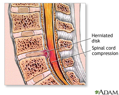
 The spine is made of bones (vertebrae) separated by soft cushions Patients usually require physical therapy to optimize spinal mobility after lumbar spine surgery. Results are variable depending on the disease treated. (intervertebral discs).
 Lumbar (lower back) spine
  disease is usually caused b
 y herniated intervertebral di The vertebrae are the bones that make up the spinal column, which surrounds and protects the spinal cord. The intervertebral disks are soft tissues that sit between each vertebrae and act as cushions between vertebrae, and absorb energy while the spinal column flexes, extends, and twists. Nerves from the spinal cord exit the spinal column between each vertebra. scs, abnormal growth of bony processes on the vertebral bodies (osteophytes), which compress spinal nerves, trauma, and narrowing (stenosis) of the spinal column around the spinal cord.
 The surgery is done while the patient is deep asleep and pain-free (general anesthesia). An incision is made over the lower back, in the midline. The vertebrae are the bones that make up the spinal column, which surrounds and protects the spinal cord. The intervertebral disks are soft tissues that sit between each vertebrae and act as cushions between vertebrae, and absorb energy while the spinal column flexes, extends, and twists. Nerves from the spinal cord exit the spinal column between each vertebra.

 The spine is made of bones (vertebrae) separated by soft cushions Patients usually require physical therapy to optimize spinal mobility after lumbar spine surgery. Results are variable depending on the disease treated. (intervertebral discs).
 Lumbar (lower back) spine
  disease is usually caused b
 y herniated intervertebral di The vertebrae are the bones that make up the spinal column, which surrounds and protects the spinal cord. The intervertebral disks are soft tissues that sit between each vertebrae and act as cushions between vertebrae, and absorb energy while the spinal column flexes, extends, and twists. Nerves from the spinal cord exit the spinal column between each vertebra. scs, abnormal growth of bony processes on the vertebral bodies (osteophytes), which compress spinal nerves, trauma, and narrowing (stenosis) of the spinal column around the spinal cord.
Symptoms of lumbar spine problems include:
- pain that extends (radiates) from the back to the buttocks or back of thigh
- pain that interferes with daily activities
- weakness of legs or feet
- numbness of legs, feet, or toes
- loss of bowel of bladder control
 The surgery is done while the patient is deep asleep and pain-free (general anesthesia). An incision is made over the lower back, in the midline. The vertebrae are the bones that make up the spinal column, which surrounds and protects the spinal cord. The intervertebral disks are soft tissues that sit between each vertebrae and act as cushions between vertebrae, and absorb energy while the spinal column flexes, extends, and twists. Nerves from the spinal cord exit the spinal column between each vertebra.
The bone that curves around and covers the spinal cord (lamina) is removed (laminectomy) and the tissue that is causing pressure on the nerve or spinal cord is removed. The hole through which the nerve passes can be enlarged to prevent further pressure on the nerve. Sometimes, a piece of bone (bone graft), interbody cages, or pedicle screws may be used to strengthen the area of surgery.

 Epidural injections involve injecting an anesthetic and an anti-inflammatory medication, such as a steroid (cortisone), near the affected nerve. This reduces the inflammation and lessens or resolves the pain. This type of epidural injection is a therapeutic one.
For diagnostic purposes, an epidural spinal injection can be done at a very specific, isolated nerve to determine if that particular nerve is the source of pain. For diagnostic purposes, only an anesthetic is injected. The immediate response to the injection is closely monitored. If the pain is completely or nearly completely relieved, then that specific nerve is the primary cause of the pain symptoms. If there is little pain relief, then another source of pain exists.
 For diagnostic purposes, facet joints can be injected in two ways: injecting anesthetic directly into the joint or anesthetizing the nerves carrying the pain signals away from the joint (medial branches of the nerve). If the majority of pain is relieved with anesthetic into the joint, then a therapeutic injection of a steroid may provide lasting neck or low back pain relief.
If anesthetic injections indicate that the nerve is the source of pain, the next step is to block the pain signals more permanently. This is done with radiofrequency ablation, or damaging the nerves that supply the joint with a "burning" technique.
This joint can also be injected for both diagnostic and therapeutic purposes. Anesthetizing the SI joint by injection under X-ray guidance is considered the gold standard for diagnosing SI joint pain. A diagnostic injection of the sacroiliac joint with anesthetic should markedly diminish the amount of pain in a specific location of the low back, buttock, or upper leg.
A therapeutic injection will typically include a steroid medication, with the goal of providing longer pain relief.
Diskography involves stimulating and "pressurizing" an intervertebral disk by injecting a liquid into the jelly-like center (nucleus pulposus) of the disk. More than one disk is injected in order to distinguish a problem disk from one without symptoms. Criteria are used to identify a painful disk by provocation discography, including the type and location of pain and the appearance of the disk on an X-ray after the procedure.
Spinal Injections
Spinal injections are used in two ways. First, they can be performed to diagnose the source of back or neck pain (diagnostic). Second, spinal injections are used as a treatment to relieve pain (therapeutic).
Most spinal injections are performed as one part of a more comprehensive treatment program. Simultaneous treatment nearly always includes an exercise program to improve or maintain spinal mobility (stretching exercises) and stability (strengthening exercises).

Spinal injections are performed under X-ray guidance, called fluoroscopy. This confirms correct placement of the medication and improves safety.
To do this, a liquid contrast (dye) is injected before the medication. If this contrast does not flow in the correct location, the needle is repositioned and additional dye is injected until the correct flow is obtained. The medication is not injected until the correct contrast flow pattern is achieved.
Epidural Injections
Epidural injections are used to treat pain that starts in the spine and radiates to an arm or leg. Arm or leg pain often occurs when a nerve is inflamed or compressed ("pinched nerve").

Epidural injection in the cervical spine.
For diagnostic purposes, an epidural spinal injection can be done at a very specific, isolated nerve to determine if that particular nerve is the source of pain. For diagnostic purposes, only an anesthetic is injected. The immediate response to the injection is closely monitored. If the pain is completely or nearly completely relieved, then that specific nerve is the primary cause of the pain symptoms. If there is little pain relief, then another source of pain exists.
Facet Joint Injections

Facet joint injection in the lumbar spine.
Facet joint injections can also be done for both diagnostic and therapeutic reasons.
These types of injections are often used when pain is caused by degenerative/arthritic conditions or injury. They are used to treat neck, middle back, or low back pain. The pain does not have to be exclusively limited to the midline spine, as these problems can cause pain to radiate into the shoulders, buttocks, or upper legs.
Facet joint injection in the cervical spine.
If anesthetic injections indicate that the nerve is the source of pain, the next step is to block the pain signals more permanently. This is done with radiofrequency ablation, or damaging the nerves that supply the joint with a "burning" technique.
Sacroiliac Joint Injections

Sacroiliac joint injection in the pelvis.
Sacroiliac joint (SI joint) injections are similar to facet joint injections in many ways. The SI joints are located between the sacrum and ilium (pelvic) bones.
Problems in the SI joints have been shown to cause pain in the low back, buttock, and leg. Typically, one joint is painful and causes pain on one side of the lower body. It is less common for both SI joints to be painful at the same time.This joint can also be injected for both diagnostic and therapeutic purposes. Anesthetizing the SI joint by injection under X-ray guidance is considered the gold standard for diagnosing SI joint pain. A diagnostic injection of the sacroiliac joint with anesthetic should markedly diminish the amount of pain in a specific location of the low back, buttock, or upper leg.
A therapeutic injection will typically include a steroid medication, with the goal of providing longer pain relief.
Provocation Diskography
Provocation diskography is a type of spine injection done only for diagnosis of pain. It does not have any pain relieving effect. In fact, it is designed to try to reproduce a person's exact or typical pain. This is to find the source of longstanding back pain that does not improve with comprehensive, conservative treatment. It can severely aggravate the existing back pain.
Diskography is performed much less commonly than the other types of spinal injections reviewed above. It is often used only if surgical treatment of low back pain is being considered. The information gained from diskography can assist in planning the surgery.Diskography involves stimulating and "pressurizing" an intervertebral disk by injecting a liquid into the jelly-like center (nucleus pulposus) of the disk. More than one disk is injected in order to distinguish a problem disk from one without symptoms. Criteria are used to identify a painful disk by provocation discography, including the type and location of pain and the appearance of the disk on an X-ray after the procedure.
Spinal injection procedures are generally safe procedures. If complications occur, they are usually mild and self-limited. The risks of spinal injections include, but are not limited to:
- Bleeding
- Infection
- Nerve injury
- Arachnoiditis
- Paralysis
- Avascular necrosis
- Spinal headache
- Muscle weakness
- Increased pain
Common side effects from steroids include:
- Facial flushing
- Increased appetite
- Menstrual irregularities
- Nausea
- Diarrhea
- Increased blood sugar
- Arthralgias
Some people are not good candidates for spinal injections. These include people with:
- Active systemic infection
- Skin infection at the site of needle puncture
- Bleeding disorder or anticoagulation
- Uncontrolled high blood pressure or diabetes
- Unstable angina or congestive heart failure
- Allergy to contrast, anesthetics, or steroids

No comments:
Post a Comment